What Is Mohs?
Mohs Micrographic Surgery was developed by Dr. Frederic Mohs in the 1930s. Dr. Mohs started developing his treatment methods on rats and other animals. He first discovered that treating cancerous tissue with zinc chloride allowed for the surgical removal and microscopic analysis of the tissue. On June 23, 1936, Dr. Mohs treated his first human patient. During the 1950s, this chemical method of cancer removal on accessible cancers transitioned to the surgical form we see today when Dr. Mohs performed a fresh tissue excision on a lower eyelid margin and had excellent results.
Mohs Surgery today is a controlled form of surgery performed by trained Dermatologists to treat skin cancer. Tissue is removed from the patient, processed through frozen sectioning, stained with Hematoxylin and Eosin Staining methods, and viewed under the microscope for diagnosis.
If the presence of skin cancer still remains, another stage of tissue specimen processing occurs until the lesion is clear of skin cancer. The most common types of cancer are Basal Cell Carcinoma (BCC) and Squamous Cell Carcinoma (SCC). Mohs Surgery provides excellent cure rates for both these types of cancers.
In more recent years, Mohs, in combination with Immunohistochemistry staining methods, has also been utilized in the removal of Melanoma, a very dangerous form of skin cancer.
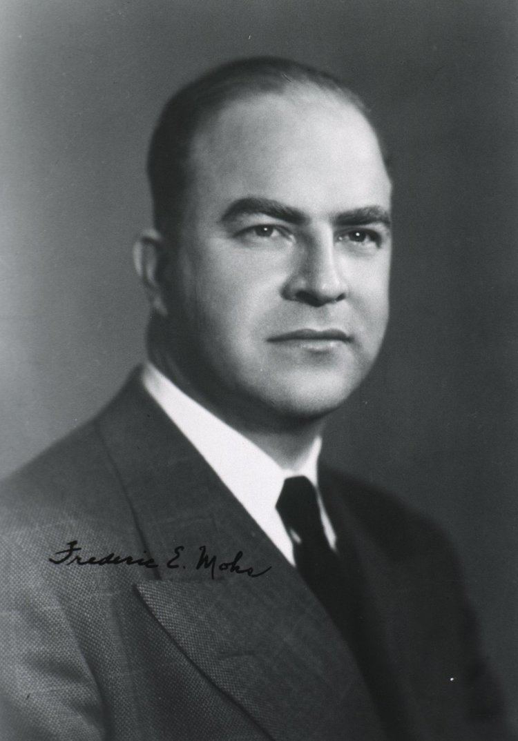
Frederic Edward Mohs, MD